The Henry Samueli School of Engineering | UC Irvine
Fluorescence lifetime imaging for extreme multiplexing
Fluorescence lifetime imaging for extreme multiplexing
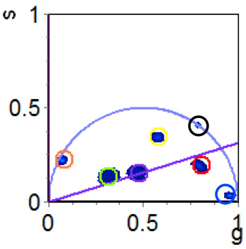
Increasing the number of detection channels that can be employed simultaneously would have an enormous impact on molecular diagnostics. This would allow more information to be obtained from a single sample, and for that information to be connected at the single cell level. The most widely used detection method in biology for multiplexing is fluorescence microscopy. Here molecules are labeled with different fluorescent probes that have unique excitation and emission wavelengths. Despite exciting developments in the fluorescent probes available, the number of detection channels is hitting an upper limit around 10. We seek to drastically extend this number by utilizing another property of fluorescent materials, the excited state lifetime. This is the time, usually on the order of tens of nanoseconds, during which fluorescent probes continue to emit light after the excitation source has been removed. Adding lifetime-based imaging to traditional fluorescence microscopy would enable expansion of the spectral-domain by the number of different probes that can be resolved in the time-domain. TWe are developing panels of nanoprobes with the same emission properties, but different fluorescence lifetimes, and testing multiplexed detection capabilities using fluorescence lifetime imaging microscopy (FLIM). This work is in collaboration with Enrico Gratton and the Laboratory for Fluorescence Dynamics (LFD) at UCI.
